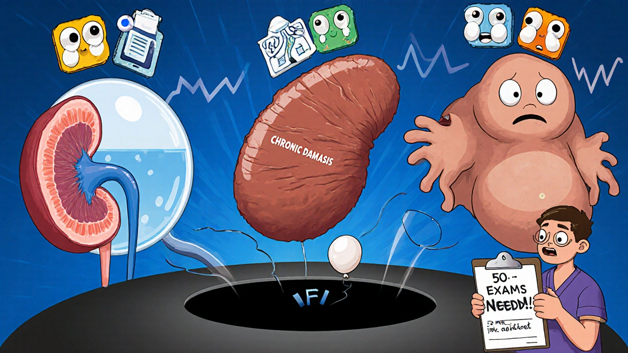
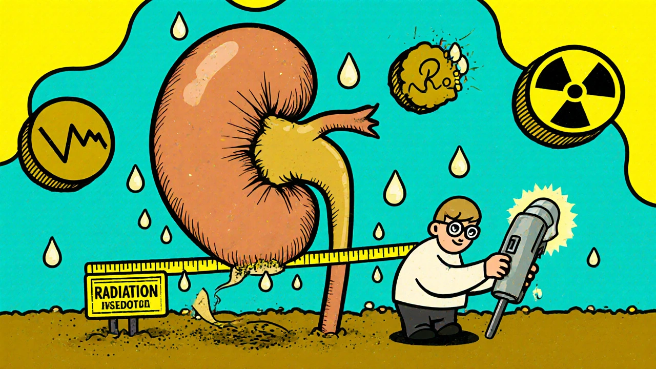
When your doctor suspects a blockage in your urinary tract or wonders if your kidneys are shrinking, they often start with one simple test: a renal ultrasound. It’s quick, safe, and doesn’t use radiation. Unlike CT scans or X-rays, which expose you to ionizing energy, ultrasound uses sound waves to create real-time images of your kidneys. This makes it the go-to first step for checking kidney size, spotting obstructions, and tracking changes over time-especially in kids, pregnant women, or anyone who needs repeated imaging.
What Renal Ultrasound Actually Shows
A renal ultrasound doesn’t just give you a blurry picture of your kidneys. It measures them. Normal adult kidneys are about 9 to 13 centimeters long. If one is significantly smaller-say, under 8 cm-it could mean long-term damage from high blood pressure, chronic infection, or scarring. Cortical thickness, the outer layer of kidney tissue, should be more than 1 centimeter. When it thins out, the kidney’s filtering power drops.
But the real power of ultrasound comes from spotting hydronephrosis. That’s the swelling inside the kidney caused by backed-up urine. Think of it like a clogged drain. When urine can’t flow out of the kidney, pressure builds, and the renal pelvis-the central collecting area-expands. Ultrasound measures this by looking at the anteroposterior diameter. A measurement over 7 millimeters in adults is a red flag. In children, even smaller amounts can signal trouble.
How Ultrasound Detects Obstruction
Obstruction isn’t always a big stone. It can be a narrowed ureter, a tumor pressing on the tube, or even a blood vessel crossing the ureteropelvic junction (UPJ). Ultrasound finds these by showing how the urine is flowing-or not flowing. The key tool here is Doppler ultrasound, which measures blood flow inside the kidney’s arteries.
One number doctors watch closely is the resistive index (RI). It’s calculated from blood flow waveforms: (peak systolic velocity minus end diastolic velocity) divided by peak systolic velocity. A normal RI is below 0.70. If it climbs to 0.70 or higher, it suggests increased resistance in the kidney’s blood vessels-often because urine pressure is squeezing them from the inside. Studies show an RI of 0.70 or more has 86.7% sensitivity and 90% specificity for detecting obstruction. That’s better than many blood tests.
Ultrasound can also spot unusual patterns. In UPJ obstruction, Doppler might show a vessel crossing the ureter, something a CT scan might miss. In severe cases, the kidney’s shape changes, the pelvis balloons, and the cortex gets compressed. These aren’t guesses-they’re measurable signs.
Why Ultrasound Beats CT for Initial Checks
CT scans are great for finding tiny stones-down to 1 or 2 millimeters. But they deliver about 10 millisieverts of radiation per scan. That’s the same as three years of natural background radiation. For someone with recurrent kidney stones, repeated CTs add up fast. Ultrasound, by contrast, uses zero radiation. That’s why emergency departments now use point-of-care ultrasound to rule out obstruction in patients with flank pain. Studies show it cuts diagnosis time by 45 minutes compared to waiting for a formal CT or ultrasound.
It’s also cheaper. In the U.S., a renal ultrasound costs between $200 and $500. A CT urogram runs $1,000 to $2,000. MR urography? That’s $1,500 to $2,500-and it’s not even good at spotting stones. Nuclear scans give functional data but need radioactive tracers and still can’t show anatomy well.
Ultrasound doesn’t need contrast dye, which can harm kidneys in people with existing damage. It’s safe for pregnant women, babies, and older adults with kidney disease. For these groups, ultrasound isn’t just preferred-it’s essential.

Where Ultrasound Falls Short
But it’s not perfect. If you’re obese-with a BMI over 35-the sound waves struggle to reach the kidneys. The images get fuzzy, and measurements become unreliable. In those cases, doctors often turn to CT or MRI, even with the radiation or cost.
Ultrasound also misses small stones. It finds about 80% of stones larger than 3 millimeters, but anything smaller? Often invisible. That’s why a negative ultrasound doesn’t always rule out a stone. If symptoms persist, a CT may still be needed.
Another problem? Operator skill. A 2018 study found up to 20% variation in kidney length measurements between inexperienced and expert sonographers. Measuring the renal pelvis correctly requires knowing exactly where to place the calipers. Getting the resistive index right means capturing clean, stable waveforms from the interlobar arteries. This isn’t something you learn in five minutes. The American College of Radiology says you need at least 50 supervised exams to become competent. Many radiology residents say it’s moderately difficult to master.
What’s New in Kidney Ultrasound
Technology is catching up. Shear-wave elastography is now being used to measure how stiff the kidney tissue is. When urine backs up, pressure builds and the kidney hardens. Studies show this stiffness increases linearly with obstruction. This could let doctors not just see blockage-but grade its severity.
Then there’s super-resolution ultrasound and ultrasound localization microscopy. These are still experimental, but they’re starting to show tiny blood vessels inside the kidney. In the future, they might detect early signs of fibrosis or reduced blood flow before the kidney even starts to swell. That’s huge. Right now, we only see damage after it happens. These tools could catch it before.
Artificial intelligence is also stepping in. Some hospitals are training AI to automatically grade hydronephrosis. Instead of relying on a radiologist’s eye, software can measure the pelvis, compare it to norms, and flag abnormalities in seconds. Early results look promising.

How the Test Is Done
There’s no special prep. You don’t need to fast. Just drink water if you’re being checked for obstruction-it helps fill the bladder and gives a clearer view. The scan takes 15 to 30 minutes. You lie on your back or side while a technician moves a handheld probe over your flank and abdomen. They’ll image both kidneys in two planes: lengthwise and crosswise.
They’ll measure:
- Kidney length, width, and thickness
- Cortical thickness
- Renal pelvis diameter
- Presence and grade of hydronephrosis (using the Society for Fetal Urology scale)
- Resistive index from at least three Doppler waveforms
They’ll also check the bladder. A full bladder can cause pressure on the ureters. An empty one might mean you’re not producing urine. Both are clues.
Who Needs This Test?
You might get a renal ultrasound if you have:
- Flank pain, especially with nausea or vomiting
- Blood in your urine without infection
- High blood pressure and shrinking kidneys
- History of kidney stones or surgery
- Abnormal kidney function on blood tests
- Pregnancy with urinary symptoms
- Children with urinary tract infections or prenatal hydronephrosis
It’s also used for follow-up. If you’ve had a kidney stone removed or surgery for UPJ obstruction, your doctor might schedule weekly ultrasounds to make sure the kidney isn’t swelling again. No radiation. No needles. Just a quick scan to keep things on track.
What Happens After the Scan
The results come back fast-often the same day. If the ultrasound shows mild hydronephrosis and normal RI, your doctor might just watch and wait. If it’s moderate to severe, they’ll likely order more tests. That could mean a CT scan to find the stone, an MRI to check for tumors, or a nuclear scan to measure kidney function.
But here’s the thing: if the ultrasound shows a clear obstruction and high RI, treatment can start right away. You might get a stent placed in the ureter to drain the kidney, or surgery to fix a narrowed UPJ. The ultrasound doesn’t just diagnose-it guides action.
And for patients who need long-term monitoring-like those with congenital blockages or recurrent stones-ultrasound is the only imaging tool that’s safe enough to use every few weeks. That’s why urologists say: "I track hydronephrosis weekly with bedside ultrasound instead of exposing them to repeated radiation."
Can a renal ultrasound detect kidney stones?
Yes, but not always. Ultrasound detects about 80% of kidney stones larger than 3 millimeters. Smaller stones, especially those under 2 mm, are often missed. CT scans are better for finding tiny stones, but ultrasound is still the first test because it’s safe, fast, and doesn’t use radiation. If the ultrasound is negative but symptoms persist, a CT may be needed to rule out a small stone.
Is renal ultrasound safe during pregnancy?
Yes, it’s the safest imaging option for pregnant women. Unlike CT or X-rays, ultrasound uses no radiation and no contrast dye. It’s routinely used to check for kidney obstruction, hydronephrosis, or infection in pregnant patients. Many women develop mild hydronephrosis during pregnancy due to hormonal changes and pressure from the growing uterus-ultrasound helps distinguish normal changes from dangerous blockages.
What does a high resistive index mean?
A resistive index (RI) of 0.70 or higher suggests increased resistance to blood flow in the kidney’s arteries, often caused by pressure from backed-up urine. It’s a strong indicator of urinary obstruction, with studies showing 86.7% sensitivity and 90% specificity. However, a high RI can also occur in kidney disease, severe dehydration, or after transplant. It’s not a diagnosis on its own-it’s a clue that needs to be interpreted with other findings like hydronephrosis and kidney size.
Can obesity affect the accuracy of a renal ultrasound?
Yes, significantly. In patients with a BMI over 35, sound waves have trouble penetrating deep enough to get clear images of the kidneys. This can make measurements of kidney size and hydronephrosis unreliable. In these cases, doctors often turn to CT or MRI, even though they involve radiation or higher cost. Obesity is one of the main reasons renal ultrasound fails to give clear answers.
How long does it take to learn to read renal ultrasounds properly?
It takes time and practice. The American College of Radiology recommends at least 50 supervised exams before a sonographer can reliably measure kidney size and resistive index. Many radiology residents say it’s moderately difficult to master. Accuracy improves with experience-novices can have up to a 20% variation in measurements compared to experts. Training programs require at least 40 supervised exams for certification, but true competence often takes more.
Do I need to prepare for a renal ultrasound?
Usually not. You don’t need to fast. But drinking water before the scan can help fill your bladder, which improves the view of the lower ureters and kidneys. If you’re being checked for obstruction, having a full bladder helps the technician see if urine is backing up. Your provider will tell you if any prep is needed, but in most cases, you can eat and drink normally.
Renal ultrasound is the quiet workhorse of kidney imaging. It doesn’t dazzle with flashy 3D images or AI-generated heat maps. But it’s reliable, safe, and deeply informative. For detecting obstruction and measuring kidney size, it’s still the best first step-and for many patients, the only one they’ll ever need.

It's fascinating how something as simple as sound waves can reveal so much about our internal health-no radiation, no needles, just pure physics doing the heavy lifting. I’ve had three renal ultrasounds in the last five years, and each time, the tech would quietly explain what they were seeing, like a gentle guide through my own body’s landscape. It’s not glamorous, but it’s deeply human. And honestly? That’s more than I can say for most of modern medicine.
As a nephrology nurse, I see this daily. Ultrasound is our first line because it’s safe and fast. But I’ve seen too many cases where techs miss hydronephrosis because they rush or don’t know where to place the calipers. Training matters. A lot. If your facility doesn’t require 50+ supervised scans before letting someone do this alone, you’re gambling with patient outcomes.
Yeah but let’s be real-this whole thing is just a glorified guess game. You measure a kidney, you see a little dilation, you panic. But what if it’s just normal variation? What if the patient drank too much coffee? The resistive index? That’s basically a number pulled out of thin air by someone who read a paper once. And don’t get me started on AI ‘grading’ hydronephrosis-how many times has a machine misread a shadow as a stone? We’re outsourcing intuition to code and calling it progress
There is, I believe, a profound elegance in the simplicity of ultrasound-it requires no exotic isotopes, no expensive infrastructure, no prolonged waiting. In rural clinics across the UK, where CT scanners are a luxury, this tool remains the quiet guardian of renal health. I’ve seen it in action: a young mother in Cornwall, pregnant, in pain, and within twenty minutes, she knew her kidneys were fine. No ionizing radiation. No fear. Just a handheld probe and a skilled hand. That is medicine at its most humane.
I work in a community clinic in Delhi where we rarely have access to CT or MRI. Ultrasound is our only reliable option, and we’ve trained our nurses to do basic renal scans with great success. The key is consistency-same machine, same protocol, same person measuring. Even with limited resources, we’ve caught early obstruction in kids with recurrent UTIs. It’s not perfect, but it’s powerful when used with care and training.
Just had my third ultrasound last week for a kidney stone I didn’t even know I had 😅 I was nervous, but the tech was so calm and explained everything. I didn’t even feel the probe! And guess what? No radiation, no needles, no weird contrast. Just me, a gel blanket, and a screen showing my own insides. It felt like magic. And now I’m drinking water like it’s my job. 💧
I appreciate the thorough breakdown. The part about operator skill stood out to me. It’s easy to assume machines or protocols are infallible, but human factors still drive accuracy. I’ve had two ultrasounds where measurements differed by nearly a centimeter-same kidney, same day, different techs. It’s humbling. Maybe we need more emphasis on standardization in training, not just tech upgrades.
This is an excellent summary of renal ultrasound applications. The emphasis on resistive index and cortical thickness is clinically sound. However, it is imperative to note that in patients with chronic kidney disease, elevated resistive indices may reflect parenchymal fibrosis rather than obstruction. Therefore, clinical correlation remains paramount. The integration of shear-wave elastography is promising, but validation in diverse populations is still ongoing.
Let’s be honest-ultrasound is the lazy man’s diagnostic tool. You want to know if there’s a stone? CT finds everything. You want to know if the kidney’s dying? A nuclear scan gives you function. Ultrasound? It’s a blurry snapshot with a 20% error rate because some tech didn’t get their 50 supervised scans. And now we’re letting AI ‘grade’ hydronephrosis? 😂 I’ve seen algorithms flag a bowel loop as hydronephrosis. That’s not innovation-that’s negligence wrapped in a fancy acronym. If you’re relying on this as your primary tool, you’re just delaying the real diagnosis.
I love that we’re still using something so simple and safe to protect people’s health. It’s like the bicycle of medical imaging-no fancy gears, no electricity, just pure, reliable motion. Every time I see someone avoid a CT scan because ultrasound caught the issue early, I feel hopeful. We don’t always need the flashiest tool-we just need the right one, used well.
Why do doctors still trust this? I’ve seen people with normal ultrasounds end up with stage 4 kidney cancer. It’s a joke. You think a blurry picture tells you everything? No. It’s just a distraction. If you’re not doing a CT first, you’re risking lives. And don’t even get me started on ‘resistive index’-that’s just fancy math to make people feel better about not using real imaging.
Ultrasound is the unsung hero of nephrology. 🏆 The fact that it can detect obstruction with 90% specificity without exposing patients to radiation is nothing short of revolutionary. And shear-wave elastography? Game-changer. We’re on the cusp of predicting fibrosis before structural damage occurs. This isn’t just imaging-it’s predictive medicine. The future is here, and it’s silent, safe, and stunningly accurate.
As a radiologist who trains residents, I can confirm: mastering renal ultrasound isn’t about equipment-it’s about pattern recognition. You learn to see the subtle asymmetry in cortical thickness, the hesitation in Doppler waveforms, the way the renal pelvis swells like a balloon under pressure. It takes years. But when you get it right, you change someone’s trajectory-maybe even save their kidney. That’s why we push for 50+ supervised cases. Because this isn’t just a scan-it’s a conversation with the body, and you have to listen carefully.